Monday, November 27, 2006

Intraepithelial lymphocytes role in preventing the leaky gut syndrome.


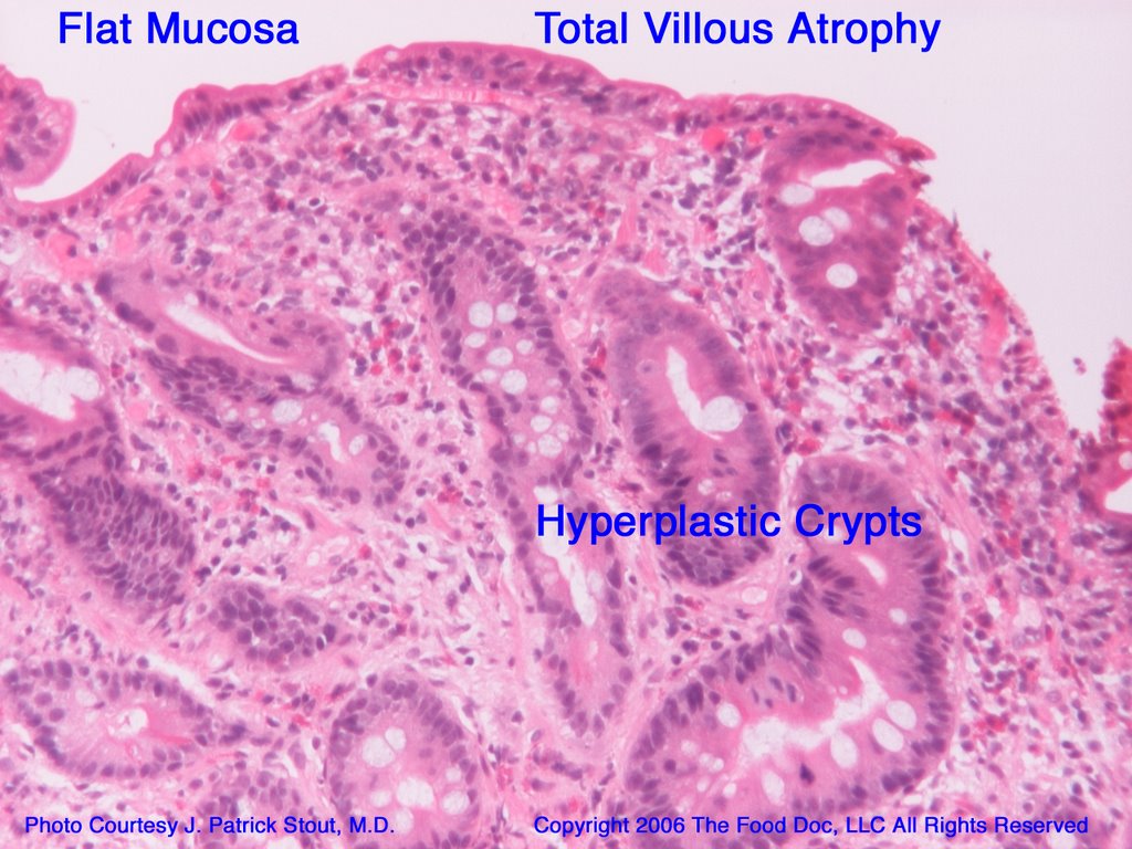
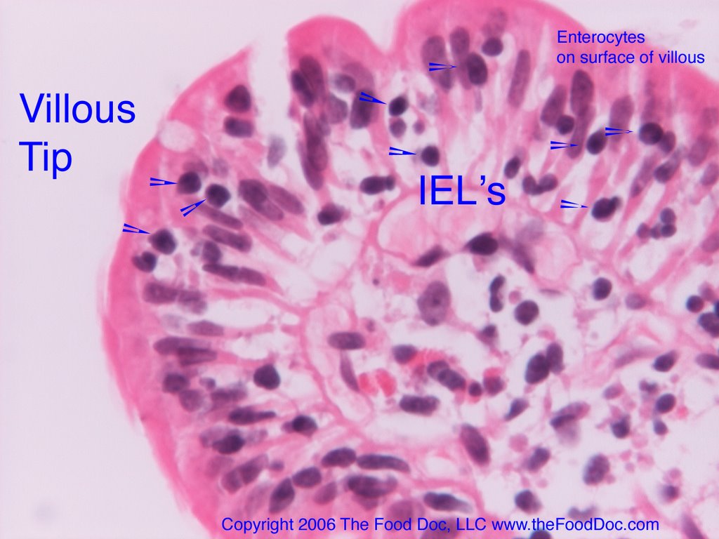
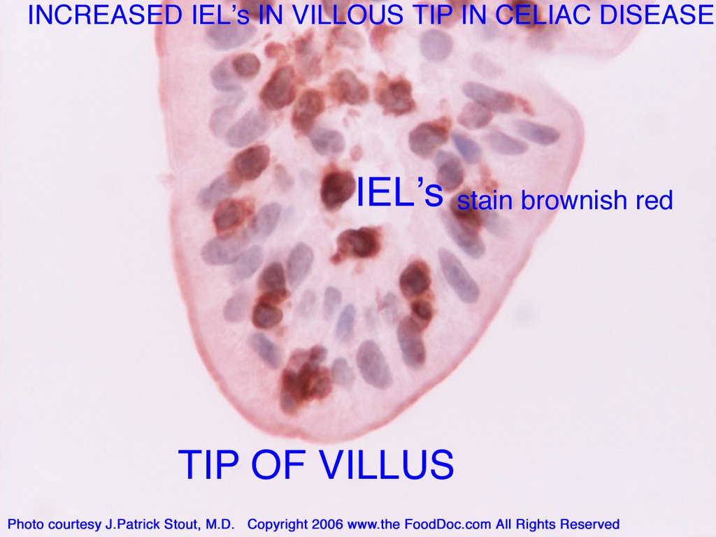
Out of the United Kingdom, comes a new article proposing that intraepithelial lymphocytes (IEL's) are important in maintaining the integrity of the tight junctions of gut to prevent the leaky gut syndrome. IEL’s are seen in increased numbers near the surface of the intestinal lining in a variety of abnormal intestinal conditions, most notably celiac disease. Being close to intestinal epithelial cells, IEL's are thought to perform several functions including tumor surveillance, restoration and repair of the lining, fighting infection, and according to new research, maintaining the tight junctions. This latter function is crucial to maintaining the gut barrier or preventing the leaky gut syndrome.

Dalton and co-investigators from the United Kingdom recently published date showing that in mice γδ+ intraepithelial lymphocytes help maintain the integrity of the epithelial cell barrier. Mice deficient in γδ+ IEL’s had severely compromised barrier function and were highly susceptible to infection from infections from Toxoplasma and Salmonella typhimurium. Reconstitution of IEL’s in deficient mice had restored intestinal barrier.

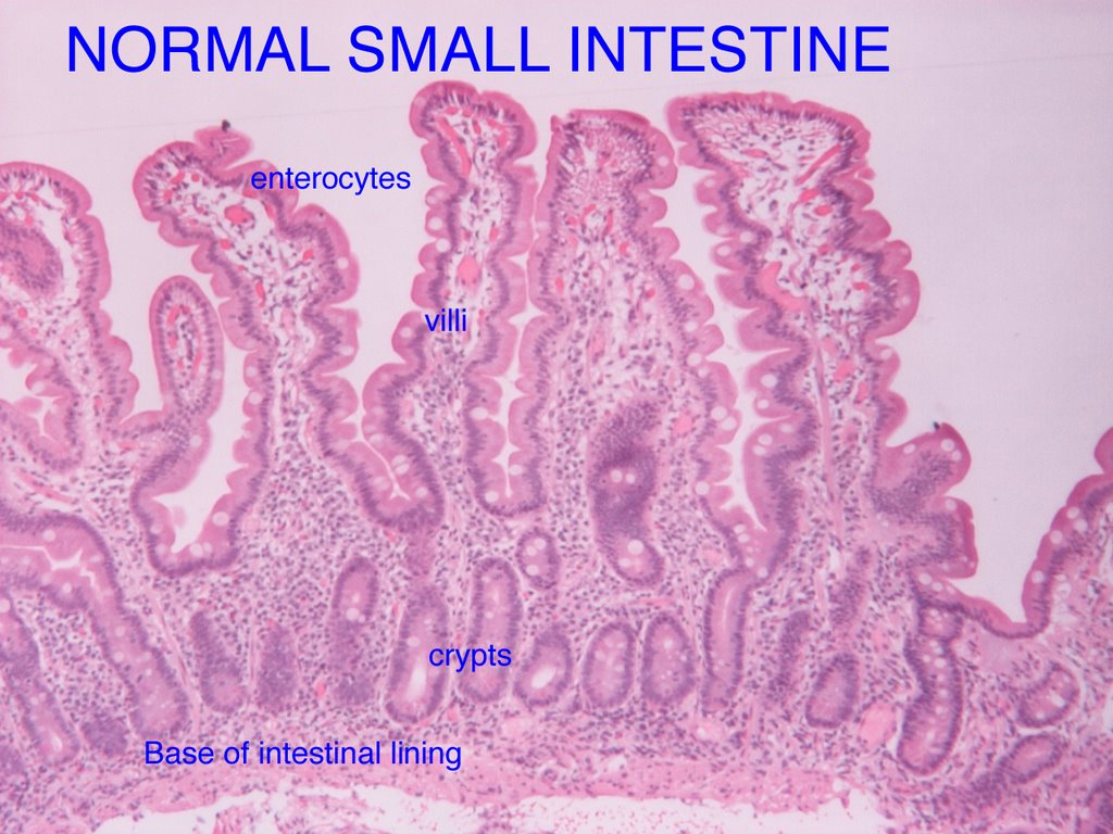
It is noteworthy that throughout the gastrointestinal tract there is only one layer of intestinal lining cells preventing what is foreign or toxic to you in what you eat or drink from gaining entry into your body. A long continuous single layer wall of epithelial cells, known as enterocytes in the small intestine, normally joined tightly, lines the entire gut. See my diagram of the intestinal villous and also my previous blogs on the intraepithelial lymphocytes. Excess IEL's are known to cause gut injury and thereby promote leaky gut, as in celiac disease.


The single layer of epithelial cells are the entry point for water and nutrients while at same time acting as a barrier to foreign proteins such as bacteria, viruses, food proteins or lectins, and noxious chemicals or toxins present in what we eat or drink. In contrast to the gut, the skin, another barrier of the body to external invaders, has a multi-layer lining of epithelial cells, rather than a single layer.

Joined by tight junctions (TJ’s) through a sophisticated system of proteins, including occludin and a regulatory protein zonulin, the epithelial gut layer serves as a barrier to foreign proteins. When this barrier is breached, foreign proteins such as bacteria and intact food proteins or lectins, may gain entry into our bodies and cause disease. Various injuries gut can result in gaps by loosening up the tight junctions (TJ’s) between the intestinal lining cells. What results is effectively a leaky gut.

How important is preventing the leaky gut syndrome? Dalton and co-workers make this statement in their discussion of their findings about the importance of IEL’s maintaining the TJ’s. “Diseases including cancer, diarrhea, inflammatory bowel disease, multiple sclerosis, diabetes, and allergic disorders are associated with disruption of TJ’s and alterations in the regulation of occludin”.

Earlier in their article they use the term “leaky” TJ complexes to describe how bacteria could infect mice deficient in the IEL’s necessary to maintain gut integrity. This article furthers our understanding on how the gut barrier is maintained and may be injured. The concept of the leaky gut as an entry way to the body for infection, toxins, and foreign food proteins resulting in infection, toxicity and/or autoimmune disease(s) continues to intrigue those of who are interested and who have eyes to see. To receive regular updates as I continue to follow important research you may subscribe to this blog by clicking on the atom feed.

Reference:
Dalton, JE et.al., Intraepithelial γδ+ lymphocytes maintain integrity of the intestinal epithelial tight junctions in response to infection. Gastroenterology 2006;131:818-829.

Copyright © 2006, The Food Doc, LLC. All Rights Reserved. www.thefooddoc.com

Wednesday, November 22, 2006

Laboratories Offering Food Intolerance Testing


Many of you have written me with questions regarding testing for food intolerance. I am listing the web links for the companies I am aware of that offer IgG tests on blood samples. These labs offer panels that test for abnormal levels of IgG antibodies for large number of foods. The testing is done on blood. Some require blood to be drawn from a vein in standard method while some offer a home test kit that employs a simple finger prick sampling returned to the laboratory.

Stool IgA antibody testing is available through Enterolab for a limited number of foods that are commonly implicated in food intolerance. Enterolab offers stool tests for gluten (gliadin, tissue transglutaminase), cow’s milk protein (casein), soy, chicken egg (ovalbumin), and Brewer’s/Baker’s dietary yeast (saccharomyces cerevisiae) by IgA method. The tests are run on stool sample provided at home and returned to Enterolab via an overnight delivery service.
Here are the web sites for the laboratories offering the above tests.

http://www.enterolab.com

http://www.greatplainslaboratory.com

http://www.optimumhealthresource.com


http://www.usbiotek.com


http://www.yorktest.com

I will review these tests in a future blog. See www.thefooddoc.com for more information including online articles.

Copyright ©2006 The Food Doc LLC, All Rights Reserved

Monday, November 20, 2006

More on the leaky gut syndrome-autoimmune disease connection.


Dr. Alessio Fasano, an expert on celiac who was one of the researchers who published the landmark article identifying the prevalence of celiac being 1:133 people in the U.S. now writes in a recent article that “the co morbidity between celiac disease and other autoimmune disorders has been clearly established.”

He reviews the two main theories that try to explain this established relationship. He concludes that the current scientific literature does not clearly explain the link but that there is “growing evidence that the loss of the intestinal barrier function typical of celiac disease could be responsible for the onset of other autoimmune diseases”.

More importantly, he theoretically proposes the autoimmune response can be stopped and “perhaps reversed if the interplay between autoimmune predisposing genes and trigger(s) is prevented or eliminated by prompt diagnosis and treatment.”

What does this mean? I believe Dr. Fasano is saying that a leaky gut exposes one’s body to food proteins, bacteria and viruses that interact with your immune system resulting in onset of various autoimmune conditions in genetically susceptible individuals. Exposure to food proteins in your gut in the setting of certain gut flora (bacteria, yeast, viruses) that your body has decided are foreign stimulates an abnormal immune response. Further, removal of certain food proteins (e.g. gluten) may not only prevent development of some autoimmune disorders but also improve or reverse some.

There is a HUGE paradigm shift showing up in the medical community concerning the role of the gut and food in health and disease.

It is my (and others) opinion that the abnormal immune responses that occur in the leaky gut syndrome from a variety of food proteins or lectins in the setting of abnormal gut flora may result in many symptoms and disorders that are too numerous to mention in this blog. However, stay tuned as I write more about this leaky gut-autoimmune connection and other food related illness and digestive conditions in the food doc blog and in online articles available at my website www.thefooddoc.com

Reference: Fasano, A. Systemic Autoimmune Disorders in Celiac Disease. Curr Opin Gastroenterol. 2006;22(6):674-679.

Saturday, November 11, 2006

The food-gut-joint axis: Foods causing arthritis


Foods cross-reacting may contribute to rheumatoid arthritis: The gut-joint axis.

Out of Norway, published in the British journal Gut, comes additional new evidence of the link between foods and rheumatoid arthritis. Professor Bradtzaeg and his colleagues at the Institute of Pathology in Oslo measured IgG, IgA, and IgM antibodies to foods in blood and intestinal fluid in 14 people with rheumatoid arthritis compared with 20 healthy people. Testing these antibodies to the following food antigens: gliadin, oats, cow’s milk proteins (casein, lactalbumin, lactoglobulin), soy, pork, cod fish, and egg (ovalbumin) revealed “particularly striking of cross reactive food antibodies in proximal gut secretions” and increased IgM antibodies to some foods in the blood but less so.

As a result measuring blood antibodies to foods in rheumatoid arthritis they believe provides little information about the role of foods whereas intestinal antibodies not only show this “striking” pattern of elevation as well as potential cumulative effect of multiple foods. The results support the connection of mucosal (gut) immune activation and causation by cross reactive foods in at least some people with rheumatoid arthritis.

What does this potentially mean? Multiple foods, especially foods that frequently show up as suspects in food allergies and sensitivities contributing to inflammatory and/or autoimmune conditions, are likely contributing to the process of gut inflammation resulting in injury (the leaky gut). This injury, especially in genetically predisposed people and in the setting of altered gut bacteria (dysbiosis), predisposes to further injury and the entry of toxic food protein-bacteria complexes resulting in inflammatory conditions such as rheumatoid arthritis.

This gut-joint axis is likely the same mechanism as the gut-brain axis (and gut-skin axis) that produces the myriad of symptoms and diseases seen that we are increasingly finding associated with food proteins and bacteria in the gut. Much more needs to learned, but it is interesting that some foods keep showing up as the usual suspects. Grains (especially wheat, barley, rye, oats, corn), dairy (casein), nightshades (potato, tomato, peppers) and peanuts, soy and other legumes particularly stand out for their proposed link to many diseases and the success of diets eliminating or restricting such foods.

The Paleolithic or Hunter-Gatherer diet specifically recommends restricting grains, dairy and legumes. Various anti-inflammatory or arthritis diets usually recommend eliminating either wheat or gluten, dairy and the nightshades. The dietary approach to autism commonly advocated is a casein-free, gluten-free diet. Despite lay public reports of great successes with such elimination diets, mainstream medicine continues to be slow to study the dietary treatment of disease. However, especially in the past two to three years more studies are appearing showing links supporting a significant role of food and bacteria in the gut and various autoimmune diseases. Continue to stay tuned for more from the Food Doc on breaking news highlighting these links. Through his soon to be launched premier website, his online articles, and his cutting edge food doc blog, Dr. Scot Lewey, the Food Doc, is dedicated to helping you eat right to feel right.

Reference: Hvatum M, Kanerud L, Hallgren, Brandtzaeg P. The gut-joint axis: cross reactive food antibodies in rheumatoid arthritis. Gut 2006; 55:1240-1247.

Copyright 2006 The Food Doc, LLC. All Rights Reserved.
www.thefooddoc.com

Thursday, November 09, 2006

The link to the leaky gut and autoimmune disease becomes clearer.


The link to the leaky gut and autoimmune disease becomes clearer.

In a just published review on alterations in intestinal permeability Dr. J. B. Medding and his colleagues in Edmonton, Alberta Canada (Arrieta, Gut, 2006) describe how the intestine serves as a barrier when normal but becomes a source of the genesis of autoimmune diseases when it becomes abnormally permeable. That is, when your gut becomes leaky (“the leaky gut syndrome”) several autoimmune diseases are known to occur.

Intestinal permeability (how leaky the gut has become) can be evaluated in a manner specific to various sites in the gut. That is, the different areas of the gut, from stomach to large intestine, can be evaluated by specific tests for leakiness and related to damage and disease in those areas. Areas of leaky gut can be observed prior to the onset of disease and appear to be involved in the development of disease, especially autoimmune diseases like Celiac disease, diabetes, Crohn’s disease, as well as probably skin diseases like atopic dermatitis, rheumatologic conditions, and even irritable bowel syndrome. The authors propose a new paradigm consisting of “three main features…

(1) A genetically susceptible immune system (the mucosal immune system), that allows the host to react abnormally to an environmental antigen.
(2) An environmental product that triggers the disease process.
(3) The ability for the environmental agent to interact with the mucosal immune system. Since the purpose of the epithelial barrier is to keep these two factors separate, and we measure this function of the barrier by permeability, the corollary of this is that an increase in permeability is a requirement for disease expression.”

What does this mean in lay terms? The gut or intestine is supposed to be a barrier to foreign proteins like foods and bacteria. If your immune system is genetically predisposed to react adversely to a certain food proteins and/or bacteria in your gut (or nerves, skin or joints) you may react with activation of damaging chemicals intended to protect you from foreign invaders that instead damage your gut making it more leaky and more vulnerable as well as your nerves, skin, and joints.

Leaky gut begets damage to your gut, nerves, skin and/or joints and you have Celiac, Crohn’s, IBS, multiple sclerosis, eczema, rheumatoid arthritis, diabetes etc. etc. Now, what is concerning for some of us interested in this area is the overload of our gut with certain foods that have proteins known as lectins that are difficult to digest and potentially toxic to the gut, especially after genetic engineering or modification. Stay tuned for more on this exciting area as I continue writing on the relationship of food, gut and disease. As your food doctor, the food doc, I hope to help you find the information you need to eat right to feel right.

Bibliography:
Arrieta MC, Bistritz L, Meddings, JB. Recent advances in clinical practice. Alterations in intestinal permeability. Gut 2006;55:1512-1520.

Copyright 2006 The Food Doc, LLC All Rights Reserved. www.thefooddoc.com

Wednesday, November 08, 2006

Food allergy, sensitivity and intolerance in irritable bowel syndrome


In the U.S., most doctors have been and continue to be skeptical that foods cause symptoms of irritable bowel syndrome (IBS) and elimination of specific foods can improve these symptoms. This is despite almost 70% of people diagnosed as having IBS report symptoms related to specific foods. There is accumulating evidence, though still criticized because of limitations of studies that make it difficult to prove, that specific foods may be the cause of symptoms in many people labeled as having IBS.

Atkinson et.al (Gut, 2004) randomized people to either an elimination diet based on elevated IgG antibody levels (YorkTest Laboratories) for specific foods or a sham diet. Those who avoided specific foods based on their IgG antibody tests had improvement in IBS symptoms (10-26% reduction) and global rating of quality of life significantly improved. Re-introducing foods for which they tested positive resulted in worsening. Zar et.al. (Am J Gastro, 2005) reported significant improvement of IBS symptoms such as pain, bloating, and alterations in bowel habits based on six month elimination of elevated food-specific IgG4 antibodies in 25 people.

Common foods reported by IBS sufferers are wheat, barley, and rye (gluten); dairy including cow's milk protein casein) and/or lactose (milk sugar); the legumes peanut and soy; yeast used to bake or brew foods; corn; shellfish and fish; nuts like almond, Brazil nut, cashew, and walnut; fruits apple, orange, and strawberry; vegetables celery, cabbage, and lettuce; the meats pork, beef, and chicken; and nightshades potato and tomato. Elimination of all of these foods in all people with IBS symptoms without specific testing or a food symptom diet diary is nearly impossible.

Interestingly, to my knowledge no one has looked at individual food-specific diet based on testing for Celiac disease, gluten intolerance or sensitivity (elevated gliadin antibodies in blood and/or stool with normal or non-diagnostic biopsy for Celiac), casein intolerance (elevated stool IgA anti-casein antibody and/or blood IgG antibody), oral allergy syndrome (OAS) history, or positive food and/or pollen allergy testing (skin prick testing, IgE RAST or CAP RAST tests, intradermal skin testing or patch skin testing). I believe it time for a more thorough evaluation for possible food cause of IBS symptoms before accepting that diagnosis and the usual non-dietary treatments.

Bibliography

Atkinson W, Sheldon TA, Shaath N, Whorwell PJ. Food elimination based on IgG antibodies in irritable bowel syndrome:a randomised controlled study. Gut 2004;53:1459-1464.

Choung RS, Talley NJ. Food Allergy and Intolerance in IBS. Gastroenterology & Hepatology October 2006;2(10):757-760.

Zar S, Benson MJ, Kumar D. Food-specific serum IgG4 and IgE titers to common food antigens in irritable bowel syndrome. American Journal Gastroenterology 2005;100:1550-1557.

Zar S, Mincher L, Benson MJ, Kumar D. Food-specific IgG4 antibody guided exclusion diet improves symptoms and rectal compliance in irritable bowel syndrome. Scandinavian Journal of Gastroenterology. 2005;40:800-807.

Copyright 2006 The Food Doc, LLC All Rights Reserved www.thefooddoc.com

Sunday, November 05, 2006

Celiac disease: What is villous atrophy and blunting?


What is villous atrophy in Celiac disease?
The Food Doc, Dr. Scot Lewey explains villous atrophy and blunting in small intestine biopsies in Celiac disease.


  • Atrophy means loss of tissue, wasting away or failure to grow.

  • The villi are often blunted or flattened due to intestinal damage or injury in severe Celiac disease.

  • The surface of the intestine will looks flat under microscope and may also visually appear so on endoscopy exam.

  • Absorption of fluids and nutrients is significantly impaired with villous atrophy. It has been described as “like trying to dry off with a tee shirt instead of a plush terry cloth bath towel.

  • Atrophy can be total (completely flat intestinal lining or total loss of villi) or subtotal (partial loss).


What is villous blunting and crypt hyperplasia in the small intestine in Celiac disease?


  • Loss of villi that is not completely flat but shortened or blunted villi are noted by an abnormal villous to crypt ratio < 3:1, meaning the height of the villi are less than three times the height of the height of the crypts (see above).

  • Crypts can enlarge (crypt hyperplasia) further making the ratio disproportionate.

  • Crypt hyperplasia with or without villous atrophy is a sign of celiac, as is the presence of intra-epitheilial lymphocytosis.


Copyright 2006, The Food Doc, LLC. All Rights Reserved. www.theFoodDoc.com

Saturday, November 04, 2006

What are intra-epithelial lymphocytes, IEL's and intra-epithelial lymphocytosis

What are intra-epithelial lymphocytes, IEL’s and intra-epithelial lymphocytosis?

The Food Doc (www.theFoodDoc.com) explains with photos intra-epithelial lymphocytosis in Celiac disease.


• Lymphocytes are a type of white blood cell important in immune function. The typically appear as purplish circles on standard H&E type stain of intestinal tissue obtained from biopsy.
• Specialized lymphocytes are present in the intestine lining.
• The intestinal lining cells are called enterocytes and are a type of epithelial cell.
• The intestinal lining is an epithelial lining.
• Lymphocytes that are activated and migrated up from crypts at the base of the intestine villi to the tip are called intra-epithelial lymphocytes.
• Normally, there are much fewer than 25-30 lymphocytes per 100 enterocytes in each villous or < 9 per villous tip.
• For thirty years, more than 40 IEL’s/100 enterocytes was considered abnormal and diagnostic of Celiac in the context of appropriate history and abnormal celiac blood tests. It is the number still used by many pathologists.
• More recently, 30 intra-epithelial lymphocytes per 100 enterocytes (6 or more IEL’s per 20 enterocytes) became the accepted criteria for intra-epithelial lymphocytosis and diagnosis of Celiac disease when villous blunting or atrophy are absent but blood tests are positive.
• Newer studies suggest that <25 IEL’s per 100 enterocytes should be the cut off for abnormal and 20-25 IEL’s/100 enterocytes is borderline and suggestive of gluten injury.

Special stains highlight IEL's in intestinal villi in Celiac disease


• IEL’s are easier to see and count with special immune chemistry stains.
• Above is a photomicrograph showing increase IEL’s in a villous tip in Celiac disease. The IEL’s are brownish red with this particular stain whereas normal intestinal cell nuclei are purplish.
• Sometimes this stain is necessary to determine IEL’s from normal intestinal nuclei and better determine the number, especially if someone has already restricted gluten in their diet.
• Studies have shown that apparently normal appearing small intestine biopsies when stained with special stains for IEL’s reveal abnormal numbers as the earliest sign of gluten injury, especially in high risk individuals like family members of people with Celiac disease.

For more information see www.theFoodDoc.com. Coming soon to www.theFoodDoc.com, a premier website with online symptom assessment tool, info wizard, online symptom diet diary, virtual office, reference material, resources, and much more. Secure online consultation now available at www.theFoodDoc.com.

Copyright © 2006 The Food Doc, LLC www.theFoodDoc.com All Rights Reserved

Above all else, guard your heart, for it affects all you do. Proverbs 4:23

Thursday, November 02, 2006

What does the normal small intestine look like under the microscope?


What does the normal small intestine look like under the microscope?

The Food Doc at www.thefooddoc.com explains and illustrates what the normal small intestine looks like under the microscope.


• Long slender villi project into the intestine opening or lumen for increased area for digestion and absorbtion of nutrients and water.
• Villi surface cells, enterocytes, are covered with microvilli that contain digestive enzymes.
• Some specialized cells, called goblet cells because they resemble a wine goblet, secrete mucus. They appear like whitish circles between the enterocytes.
• At the base of the villi are crypts, collections of cells from which new lining cells arise and migrate up from to replace old or injured enterocytes and goblet cells.
• The villi are normally 3-5 times the length of height of crypts, i.e. villous-crypt ratio is normally >3-5:1 but is reduced when the inestinal lining is damaged. This results in blunting or flattening of villi and can be compared to bare spots in a terry cloth towel where absorption is impaired.
• Mixed within the intestinal lining are white blood cells known as lymphocytes. They are activated in response to injury or attack and migrate to tips of villi where they are known as intra-epithelial lymphocytes (IEL’s) and there abnormal presence is known as intra-epithelial lymphocytosis. This is the earliest feature of Celiac disease though not specific for Celiac as some other conditions can cause intra-epithelial lymphocytosis.

For more information visit www.theFoodDoc.com
Visit the Virtual Practice of Dr. Scot Lewey on HealthTap