What is villous atrophy in Celiac disease?
The Food Doc, Dr. Scot Lewey explains villous atrophy and blunting in small intestine biopsies in Celiac disease.
- Atrophy means loss of tissue, wasting away or failure to grow.
- The villi are often blunted or flattened due to intestinal damage or injury in severe Celiac disease.
- The surface of the intestine will looks flat under microscope and may also visually appear so on endoscopy exam.
- Absorption of fluids and nutrients is significantly impaired with villous atrophy. It has been described as “like trying to dry off with a tee shirt instead of a plush terry cloth bath towel.
- Atrophy can be total (completely flat intestinal lining or total loss of villi) or subtotal (partial loss).

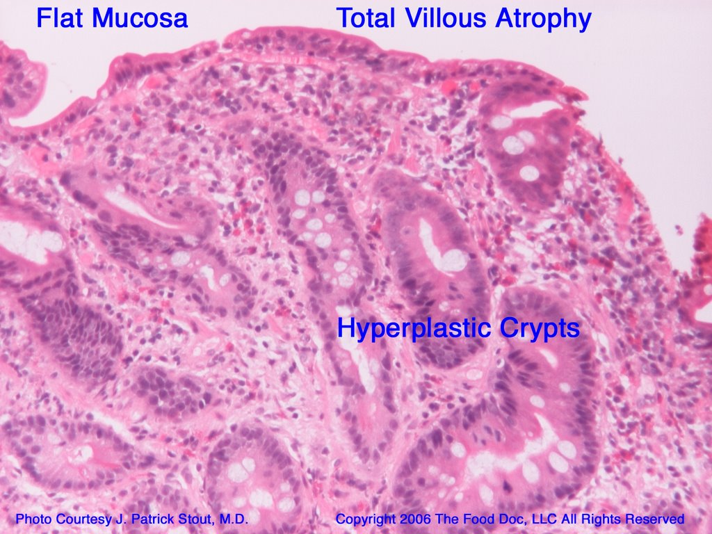
What is villous blunting and crypt hyperplasia in the small intestine in Celiac disease?
- Loss of villi that is not completely flat but shortened or blunted villi are noted by an abnormal villous to crypt ratio < 3:1, meaning the height of the villi are less than three times the height of the height of the crypts (see above).
- Crypts can enlarge (crypt hyperplasia) further making the ratio disproportionate.
- Crypt hyperplasia with or without villous atrophy is a sign of celiac, as is the presence of intra-epitheilial lymphocytosis.
Copyright 2006, The Food Doc, LLC. All Rights Reserved. www.theFoodDoc.com