What does the normal small intestine look like under the microscope?
The Food Doc at www.thefooddoc.com explains and illustrates what the normal small intestine looks like under the microscope.
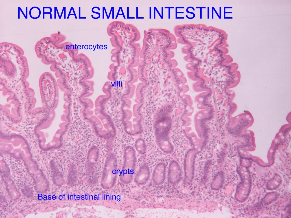
• Long slender villi project into the intestine opening or lumen for increased area for digestion and absorbtion of nutrients and water.
• Villi surface cells, enterocytes, are covered with microvilli that contain digestive enzymes.
• Some specialized cells, called goblet cells because they resemble a wine goblet, secrete mucus. They appear like whitish circles between the enterocytes.
• At the base of the villi are crypts, collections of cells from which new lining cells arise and migrate up from to replace old or injured enterocytes and goblet cells.
• The villi are normally 3-5 times the length of height of crypts, i.e. villous-crypt ratio is normally >3-5:1 but is reduced when the inestinal lining is damaged. This results in blunting or flattening of villi and can be compared to bare spots in a terry cloth towel where absorption is impaired.
• Mixed within the intestinal lining are white blood cells known as lymphocytes. They are activated in response to injury or attack and migrate to tips of villi where they are known as intra-epithelial lymphocytes (IEL’s) and there abnormal presence is known as intra-epithelial lymphocytosis. This is the earliest feature of Celiac disease though not specific for Celiac as some other conditions can cause intra-epithelial lymphocytosis.
For more information visit www.theFoodDoc.com